Rigorous research.
Real-world impact.

Behind Melanoma Score is a research programme led by Professor Gustav Stålhammar at St. Erik Eye Hospital and Karolinska Institutet. Here is how the model was built and validated, and what it means for patients, healthcare providers, and optometrists.

  1. Find the lesion
  2. Score from 0 to 1
Figure 1. Simplified from the method in Ophthalmology Science, 2024, where 0.63 was the operating point, as in TVST, 2025. The lesion is painted, not a patient's, so no score is shown.

Figure 1, an illustration. An image of the back of the eye, with a painted pigmented lesion. A circle centres on the lesion, and the model's method, as described in Ophthalmology Science, 2024 and simplified here to two steps, plays out. First, an outline is drawn around the lesion and the circle becomes a square centred on it. Second, the outline unrolls into a scale from 0 to 1 under the square, with a mark at 0.63, the operating point used in the Ophthalmology Science 2024 and TVST 2025 studies. No score is shown, because the lesion is painted and not a patient's.

At a glance

The short version, in nine lines.

Each line links to its section. Every figure names its source.

Developed by
Prof. Gustav Stålhammar's research group at St. Erik Eye Hospital and Karolinska Institutet, with Mats Holmström. Ten authors across three papers.
Intended use
Decision support for clinicians who assess pigmented lesions at the back of the eye, from opticians and optometrists to ocular oncologists. Cancers, 2026
Input
One colour photograph of the back of the eye. The studies used standard field and ultra-widefield photographs from two camera systems. Ophthalmology Science, 2024
Output
A risk score from 0 to 1. The same image gives the same score every time, whoever runs it. Cancers, 2026
Data
In the 2024 study, 802 photographs from 688 patients, each assessed by an ocular oncologist. Harmless meant at least five years without being rediagnosed as melanoma. Ophthalmology Science, 2024
Tested
Three peer-reviewed studies: against twelve ophthalmologists (Ophthalmology Science, 2024), 29 opticians and optometrists on 25 photographs with five melanomas (TVST, 2025), and the MOLES checklist in expert hands (Cancers, 2026).
Key result
In the 2024 study, AUC 0.88 on both test sets. At the 0.63 threshold, 100 % sensitivity with 74 % specificity (internal set) and 80 % with 81 % (external set). Ophthalmology Science, 2024
Still open
Tested on images from several hospitals and validated on an external test set. Prospective multicentre studies are planned.
Disclosures
Each paper names its funding. The 2026 paper declares that Mats Holmström and Gustav Stålhammar are founders of Eyedentity AB and hold equity in the company. Cancers, 2026

01Studies

Developed, compared, and tested where referrals begin.

From development and validation, to optician triage, to a head-to-head comparison with the MOLES checklist.

Study 1, Ophthalmology Science, 2024: Can the model tell a small melanoma from a harmless lesion?

A retrospective multicentre study of 802 photographs from 688 patients, all examined at St. Erik Eye Hospital. The model was built on one part of them and tested on two sets it had never seen: 86 photographs held back from the same collection, and 53 taken at other clinics across Sweden before the patients came to St. Erik. Twelve ophthalmologists, from residents to ocular oncologists, read the same 86 photographs without knowing the answers.

AUC, how often a score ranks a melanoma above a harmless lesion: 0.5 is a coin toss, 1.0 is perfect. Whiskers show 95 % confidence intervals. Values from the paper's Figure 4 and its results.Ophthalmology Science, 2024
Figure 2. AUC on the two test sets
Test setMethodAUC95 % confidence interval
Internal test set, 86 photographs, held back from the same collectionThe model, from the photograph alone0.880.82 to 0.95
Internal test set, 86 photographs, held back from the same collectionMOLES, Checklists, also scored with ultrasound or OCT0.770.66 to 0.88
Internal test set, 86 photographs, held back from the same collectionTFSOM-UHHD, Checklists, also scored with ultrasound or OCT0.670.54 to 0.81
Internal test set, 86 photographs, held back from the same collectionBoth checklists were scored by ophthalmologists who also had ultrasound or OCT from the records. The model had the photograph only.
External test set, 53 photographs, other clinics in SwedenThe model, from the photograph alone0.880.74 to 1.00
External test set, 53 photographs, other clinics in SwedenThe ophthalmologists read the internal set only, and the checklists were compared with the model on the internal set only.

At the 0.63 threshold, set before testing, the model had 100 % sensitivity and 74 % specificity on the internal set, and 80 % sensitivity and 81 % specificity on the external set. On the internal set, the one the twelve ophthalmologists also read, its sensitivity was significantly higher than the residents' and the consultants' and not significantly different from the ocular oncologists'; its specificity did not differ significantly from any group's. Ophthalmology Science, 2024

The authors' conclusion

“This study develops and validates a deep learning algorithm for differentiating small choroidal melanomas from nevi, matching or surpassing the discriminatory performance of experienced human ophthalmologists.”

Ophthalmology Science, 2024

The authors' caveat

“Further research will aim to validate its utility in clinical settings.”

Ophthalmology Science, 2024

Study 2, TVST, 2025: How do opticians triage small pigmented lesions, and how does the model's threshold compare on the same images?

Twenty-nine Swedish opticians and optometrists graded 25 photographs of small pigmented lesions, five of them melanomas. They scored each one with MOLES, the checklist the Swedish referral guideline uses, and decided whether to refer. The study then compares their referrals with the model's threshold on the same images.

Opticians: the referrals they recorded, 29 decisions per image. Model: its own classification of each image at the 0.63 threshold, calculated on the same images. The opticians' own decisions after seeing the score were not recorded.TVST, 2025
Figure 3. Share referred, same 25 images
LesionsOpticians or modelShare referred95 % confidence interval (not reported per group)
Harmless lesions, 20 imagesThe model's threshold, calculated on the same images10 %
Harmless lesions, 20 imagesOpticians, the referrals they recorded68 %
Harmless lesions, 20 imagesThe opticians' 580 decisions included 397 referrals; the model's threshold flagged 2 of the 20 images.
Melanomas, 5 imagesThe model's threshold, calculated on the same images80 %
Melanomas, 5 imagesOpticians, the referrals they recorded95 %
Melanomas, 5 imagesThe opticians' 145 decisions included 138 referrals; the model's threshold flagged 4 of the 5 images. It missed one of the five.

In the authors' words, the opticians' low specificity “reflects guideline structure rather than clinical incompetence”: the guideline asks them to refer at a MOLES score of 1 or more, and a score of 1 is what MOLES itself calls a low-risk nevus. The authors write that the model's threshold on the same images would have “reduced false-positive referrals 10-fold”. With five melanomas in the set, that came at the cost of one: the threshold flagged 4 of 5, where the opticians' recorded decisions referred 95 %. TVST, 2025

From the opticians' questionnaire

In the study's first step, 54 opticians and optometrists from 17 shops answered a questionnaire; 29 graded the images in the second.

  • 83 %find it hard to judge whether the referral criteria are metn = 54
  • 65 %typically take two minutes or more to assess a lesionn = 54
  • 70 %see five or more customers with pigmented lesions a yearn = 54
  • 85 %have a fundus camera where they workn = 54

TVST, 2025

The authors' conclusion

“Swedish opticians and optometrists detect small pigmented choroidal lesions with high sensitivity but poor specificity, leading to frequent, unnecessary referrals.”

TVST, 2025

The authors' caveat

“Notably, among these 25 lesions, only 5 were melanomas, meaning that a single missed melanoma reduced sensitivity from 100 % to 80 %.”

TVST, 2025

Study 3, Cancers, 2026: How does the model's score compare with the MOLES checklist in expert hands?

Eighty-six lesions from St. Erik Eye Hospital, 29 of them melanomas: the 2024 study's internal test set, none of it used to build the model. Ocular oncologists scored MOLES with OCT and autofluorescence at hand, the checklist's best case. The model, which the paper calls “MelAInoma”, had the colour photograph only. No threshold was applied; the score was used on its full scale from 0 to 1.

The differences between the two were “not statistically detectable”, in the authors' words. Whiskers show 95 % confidence intervals.Cancers, 2026
Figure 4. AUC by camera type
CameraMethodAUC95 % confidence interval
Standard field, 45°, 55 photographsThe model, from the photograph alone0.870.78 to 0.96
Standard field, 45°, 55 photographsMOLES, scored by ocular oncologists with OCT and autofluorescence0.800.67 to 0.93
Standard field, 45°, 55 photographsP = 0.42 for the difference (DeLong test)
Ultra-widefield, 200°, 31 photographsThe model, from the photograph alone0.870.72 to 1.00
Ultra-widefield, 200°, 31 photographsMOLES, scored by ocular oncologists with OCT and autofluorescence0.790.61 to 0.97
Ultra-widefield, 200°, 31 photographsP = 0.48 for the difference (DeLong test)

The model's score explained more of the variation in diagnosis than MOLES did, Tjur R² 0.38 against 0.27. The two scores share only 16 % of their variance, so they often rate the same lesion differently. Cancers, 2026

The authors' conclusion

“MelAInoma demonstrates slightly stronger association with melanoma diagnosis and provides fully reproducible output, supporting its role as a complementary aid in lesion triage.”

Cancers, 2026

The authors' caveat

“Statistical association does not by itself establish clinical utility, which depends on referral thresholds, decision context, and acceptable trade-offs between sensitivity and specificity.”

Cancers, 2026

Conflict of interest, as declared in the paper

“Mats Holmström and Gustav Stålhammar are founders of Eyedentity AB and hold equity in the company, which develops the MelAInoma software evaluated in this study. The remaining authors declare that the research was conducted in the absence of any commercial or financial relationships that could be construed as a potential conflict of interest.”

Cancers, 2026

02Method

The model finds the lesion first, then scores it.

Here is how the model in Figure 1 gets from photograph to answer, as the 2024 paper describes it.

  1. Find the lesion

    The model marks the pixels in the photograph that belong to the lesion.

  2. Crop around it

    It centres a square crop on the lesion, so the next step reads the lesion rather than the whole retina.

  3. Score from 0 to 1

    It reads the crop and returns one risk score between 0 and 1. A threshold turns the score into a recommendation: refer, or do not refer. The 2024 study chose one threshold, 0.63, on the training and tuning images and kept it unchanged for both test sets. The 2025 study used the same threshold.

In the 2026 paper's words, the model “provides fully reproducible output, yielding the same score each time a given image is analyzed, independent of the user”

Cancers, 2026

Every label came from a specialist's examination.

The 2024 study drew on photographs from multiple institutions across Sweden, collected at St. Erik Eye Hospital's ocular oncology service, the national referral centre where every patient was examined.

Figure 5. How the 2024 study's 802 photographs were split

  1. Training495 images, 410 patients
  2. Tuning168 images, 139 patients
  3. Internal test86 images, 86 patients
  4. External test53 images, 53 patients, other clinics
No image was used in more than one set, and no patient crossed between sets. The threshold was chosen on the training and tuning sets (the paper calls the tuning set the validation set); the two test sets were only used to test.Ophthalmology Science, 2024

How each image got its answer

  • Every patient was examined by an ocular oncologist, with photographs, OCT, ultrasound and slit lamp, and a biopsy in the rare case where the examinations were inconclusive.
  • A lesion counted as harmless only after at least five years without being rediagnosed as melanoma.
  • A lesion counted as melanoma if it was diagnosed as one, then or later, for example after it grew. The authors added this rule to help the model pick up early signs of malignancy, at a time when a small melanoma is hard to tell from a harmless lesion.

Ophthalmology Science, 2024

03Intended use

One question, asked of one photograph.

Is a pigmented lesion at the back of the eye a harmless nevus, or a small melanoma? The model gives a risk score for that one question, from a colour photograph.

Such lesions are common. Up to 700,000 people in Sweden have one, according to SVT. Almost all stay harmless, but a small melanoma can look just like one.

Telling them apart is hard even for experienced ocular oncologists, and a growing number are found by chance: in diabetic eye screening, before cataract surgery, and in the fundus photographs opticians and optometrists take, as the opticians' study in TVST (2025) describes.

4 % to 7 % of Caucasian adults have a choroidal nevus, according to the population studies the 2024 paper cites. Among people of African and Asian descent it is under 1 %.

Ophthalmology Science, 2024

“The intended use is decision support for clinicians who assess pigmented choroidal lesions with varying frequency and expertise, including opticians, optometrists, general ophthalmologists, and subspecialized ocular oncologists.”

Cancers, 2026

A risk assessment that supports a referral decision. The clinician decides.

04Limits

What the studies leave open.

Every paper names its own limits. Here they are side by side, each with the authors' own words.

Referred patients

Every patient was examined at one national referral centre, so the mix of cases is a specialist's, not an optician's. Only the 2024 study had a test set from other clinics; the 2026 paper names its single centre as a limit.

“conducted at a single national ocular oncology referral center, which may limit generalizability” Cancers, 2026

Two cameras

Only two kinds of camera were used, one standard field and one ultra-widefield.

“we advise against its application without prior adaptation and revalidation on other types of images” Ophthalmology Science, 2024

Small sets

The test sets are small, and the opticians' study held five melanomas.

“a single missed melanoma reduced sensitivity from 100 % to 80 %” TVST, 2025

Clinical answers

The reference answers came from ocular oncologists' clinical assessment, with a biopsy only in rare unclear cases. In the 2026 study, no lesion was confirmed by tissue.

“none of the included lesions underwent histopathologic confirmation” Cancers, 2026

Past photographs

All three studies used photographs taken in the past, and none recorded a decision made with the score in hand.

“Further investigation is needed, ideally in prospective cohorts, to validate the practical application of this algorithm in clinical settings” Ophthalmology Science, 2024

05People

The people behind the evidence.

Prof. Gustav Stålhammar

  • Professor of ophthalmology at Karolinska Institutet
  • Senior consultant and ocular oncologist at St. Erik Eye Hospital

He leads the research behind the model and is the corresponding author on all three papers. At St. Erik Eye Hospital he is one of a handful of specialists who treat the whole country's cases of uveal melanoma. He is also a founder of Eyedentity and its Chief Scientific Officer.

100+
peer-reviewed articles by Prof. Stålhammar, three of them on the melanoma model
#4
in the world in uveal melanoma research over the past five years, ranked by ScholarGPSSee the ranking (opens in a new tab)
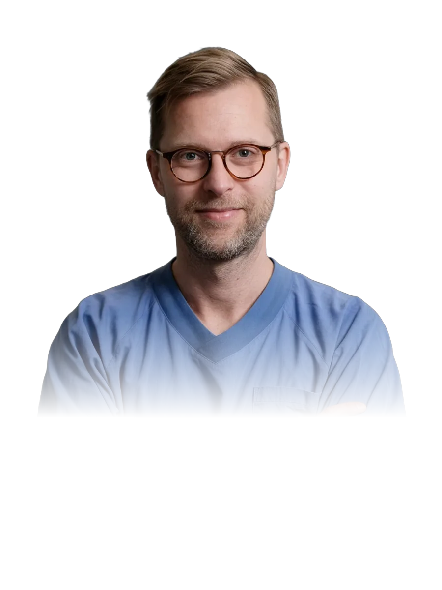
Prof. Gustav Stålhammar in blue scrubs, arms crossed

Authors on the three papers

Most of the authors are based at St. Erik Eye Hospital, in its Ocular Oncology Service and Ophthalmic Pathology Laboratory, and at Karolinska Institutet's Division of Eye and Vision, with co-authors from Kalmar County Hospital on the 2024 paper, Synologen AB on the 2025 paper and Eyedentity AB on the 2026 paper. The first studies on the model were published before the company existed.

06Papers

Read the papers in full.

All three are open access under CC BY 4.0. The TVST and Cancers titles use the model's research name; the Ophthalmology Science paper does not name it.

  • Open access

    Development and Validation of a Deep Learning Algorithm for Differentiation of Choroidal Nevi from Small Melanoma in Fundus Photographs

    Funding
    “Support for this study was provided to G.S. from the Swedish Society of Medicine (SLS-971390), Region Stockholm (FoUI-981345), the Swedish Cancer Society (23 2613 Fk), and the Swedish Eye Foundation (2023-05-04). The sponsors or funding organizations had no role in the design or conduct of this study.”
    Ethics
    Swedish Ethical Review Authority, reference 2022-06210-02. Informed consent was waived because the study was retrospective.
    Conflicts
    “All authors have completed and submitted the ICMJE disclosures form.” The paper's disclosure section lists the grant support above and nothing else.

  • Open access

    Challenges for Opticians in Evaluating Small Pigmented Choroidal Lesions: Potential Support From the MelAInoma Deep Learning Algorithm

    Funding
    “Supported by the Swedish Eye Foundation (Ögonfonden, 23-05-04; GS). The sponsor or funding organization had no role in the design or conduct of this study.”
    Ethics
    Swedish Ethical Review Authority, reference 2024-02620-02. Informed consent was waived because the study was retrospective.
    Conflicts
    “Disclosure: S. Sabazade, None; M.A. Lumia Michalski, None; S. Wittskog, None; M. Holmström, None; M. Nilsson, None; G. Stålhammar, None”

  • Open access

    Comparison of MOLES and MelAInoma for Differentiating Small Choroidal Melanomas from Nevi

    Funding
    “This research was funded by Karolinska Institutet, grant number 2023-02622.”
    Ethics
    Swedish Ethical Review Authority, reference 2025-05054-01. Patient consent was waived because the study was retrospective.
    Conflicts
    “Mats Holmström and Gustav Stålhammar are founders of Eyedentity AB and hold equity in the company, which develops the MelAInoma software evaluated in this study. The remaining authors declare that the research was conducted in the absence of any commercial or financial relationships that could be construed as a potential conflict of interest.”

Contact us