Melanoma Score.
Seen in seconds.

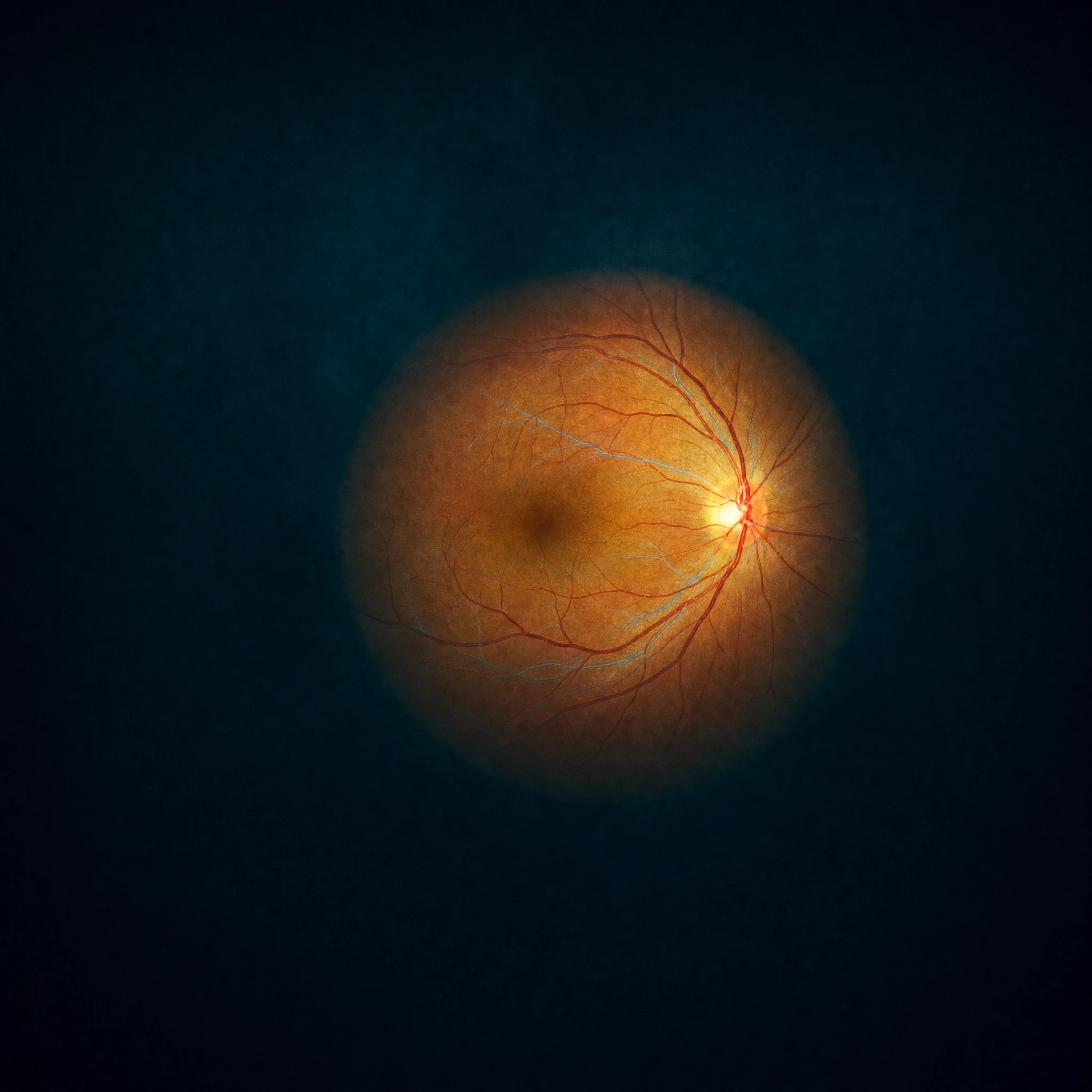
Seconds after the routine fundus photo, the optician has a risk assessment for uveal melanoma. Same camera, same visit, nothing new to do.

A pigmented lesion is common.
The hard part is telling which is which.

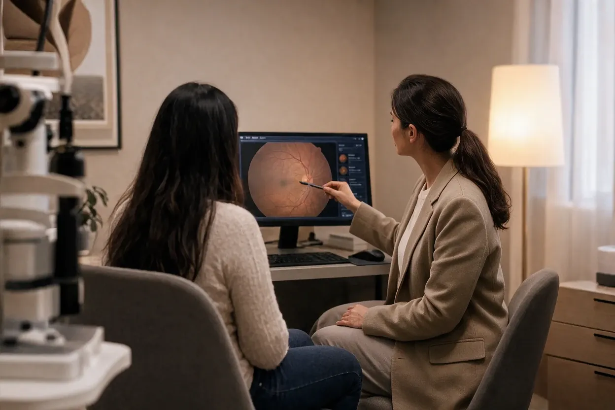
An optician pointing at the fundus image on the screen, the patient beside her

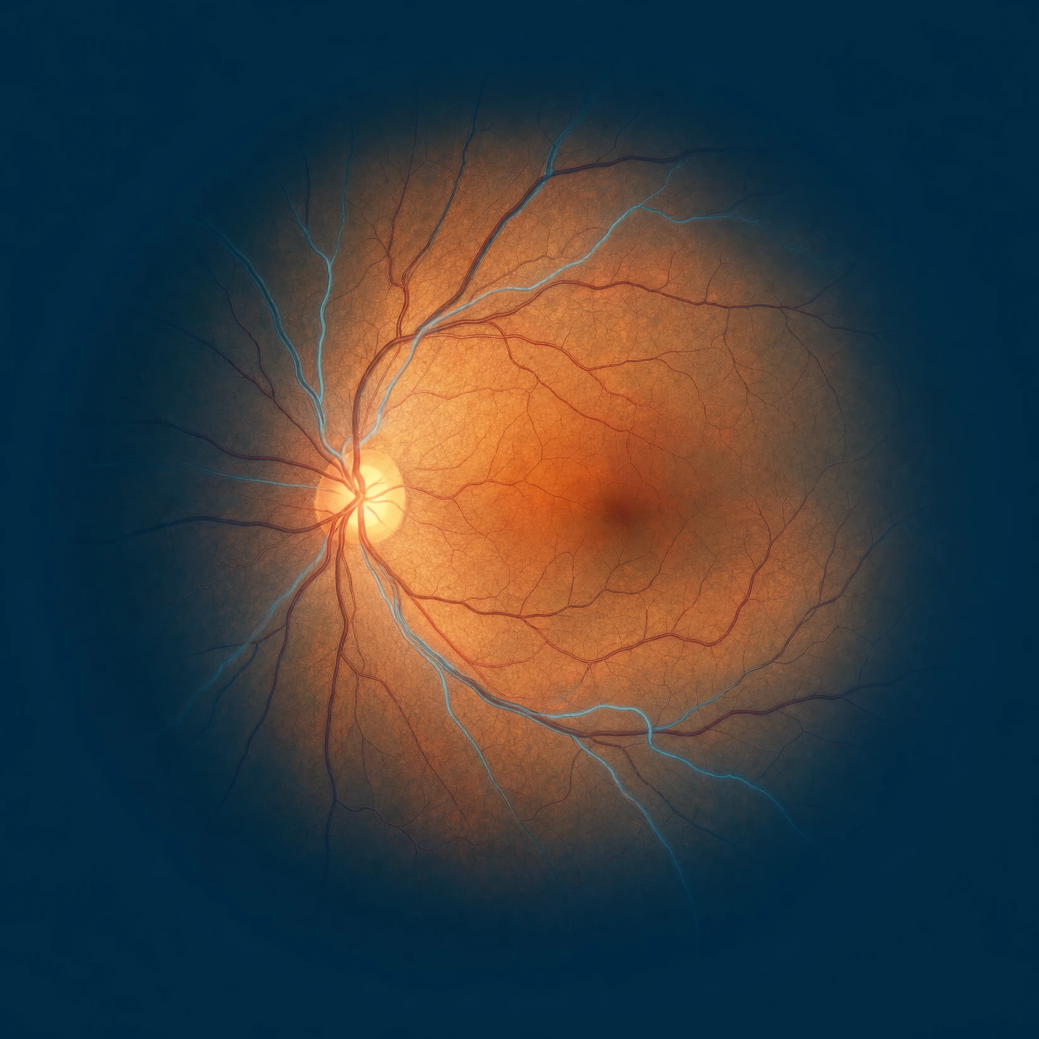
Opticians in Scandinavia take about 40,000 fundus images a day. Up to 700,000 people in Sweden have a pigmented lesion in the back of the eye. Almost all of them are harmless, a few are an aggressive melanoma. So an uncertain lesion is passed on: to a colleague, or to an external second opinion for a fee, and the answer comes back days later, when the customer has already left.

It is notoriously difficult to assess the spot with the naked eye.

Prof. Gustav Stålhammar, to SVT Nyheter, April 2026
SVT NyheterWatch the feature, Opens in a new tab

What the optician sees: an answer, not just a number.

The optician showing the retinal image on a tablet across the desk

Seconds after the photo is taken, the result is back in the room, clear and ready for the optician to act on before the customer leaves.

Score

A risk assessment of the pigmented lesion, read from the routine photo. The same image always gives the same score.

Recommendation

What the result means and what to do next, so the optician can decide while the customer is still in the chair.

Image quality

The recommendation includes an image quality check, so the user can take the photo quality into account and retake the image if needed.

Built where the disease is treated.

Melanoma Score is a machine learning model, trained on longitudinal clinical data in partnership with St. Erik Eye Hospital, where a handful of specialists diagnose and treat uveal melanoma for the whole country. The training images come from patients examined by those specialists, and a lesion counts as benign only after five years without a change of diagnosis. The model is trained and fixed: one model for one question. Behind it are Prof. Gustav Stålhammar, who leads the research at Karolinska Institutet and St. Erik, and Mats Holmström, who brought the world's first machine learning based automated radiation planning product to market at RaySearch.

See the research

What the model rests on:

  • Ophthalmology Science, 2024

    Developed and validated on fundus photographs from clinics nationwide. The model matched or surpassed experienced ophthalmologists at telling small melanomas from harmless pigmented lesions in the same photographs.

  • Translational Vision Science & Technology, 2025

    Opticians graded a set of small pigmented lesions by the referral guideline and referred most of the harmless ones. On the same images, the model's threshold would have cut false-positive referrals to a tenth, in the authors' words.

  • Cancers, 2026

    Compared with the MOLES scoring system, the model's score tracked the diagnosis more closely, and the same image always gives the same score.

Prof. Gustav Stålhammar, who leads the research, is ranked fourth in the world in uveal melanoma research over the past five years (ScholarGPS).

Bringing specialist expertise to every eye exam.

The facts, in one place.

For the clinical lead and the IT lead:

Reads
Colour fundus photographs from the cameras already in your stores, with a model that is trained and fixed.
Returns
A risk level for suspected uveal melanoma, a recommendation, and a note on image quality. In seconds.
Needs
The camera and the exam you already have. No new equipment, no new task, no new infrastructure. GDPR compliant, built to the requirements of medical software.
Stores
Nothing. The image is uploaded, analysed and discarded, and Eyedentity keeps no patient register.
Records
The referral text is written for the chain's records system, so nothing is typed twice.

Whether you are an eye clinic, hospital or optician, contact us to explore how Eyedentity can support your organisation.

Contact us